
Dry eye syndrome ( Xerophthalmia )
Dry eye syndrome remains a disorder of deficient wetness of the cornea and conjunctiva due to a violation of the quality and quantity of lacrimal fluid and instability of the lacrimal film. Manifestations of dry eye syndrome include burning and stinging a feeling of sand in the eyes, lacrimation, photophobia, rapid fatigue during visual work, intolerance to dry and dusty air. Dry eye syndrome is diagnosed based on the results of biomicroscopy, Schirmer and Norn tests, fluorescein instillation test, tiascopy, osmometry, crystallography of lacrimal fluid, cytological examination of a smear from the conjunctiva. Artificial tear preparations, obturation of the lacrimal ducts, tarsorrhaphy, keratoplasty, and salivary gland transplantation are indicated as treatment for dry eye syndrome.

General information
Dry eye syndrome is experienced by a lot of people in ophthalmology , with the eyes feeling dry and signs of xerosis usually developing. Nine to eighteen percent of the population have dry eye syndrome and most affected individuals are women, accounting for almost three out of four dry eye cases. The disease is more common as individuals increase in age: the percentage of people with dry eye increases from 12% before age 50 to 67% for those 50 and over.
Usually, a layer of tears (about 10 µm thick) covers the front surface of the eye. The outer lipid layer an greasy secretion of the meibomian glands confirms the gliding of the greater eyelid on the surface of the watch and slows down the fading of the tear film. The aqueous layer with dissolved electrolytes and organic compounds washes out foreign bodies from the eye , provides the cornea with nutrients and oxygen, and creates immune protection. The mucin layer a mucous secretion of goblet and epithelial cells directly contacts the cornea:
makes its surface even and smooth binding the tear film to this one and ensuring high quality of vision.
Approximately every 10 seconds the tear film ruptures initiating blinking of the eyelids and renewal of the tear fluid restoring its integrity. Violation of the stability of the precorneal tear film leads to its frequent ruptures, dryness of the corneal surface and conjunctiva, peters anormaly and the development of dry eye syndrome.
Reasons
Dry eye syndrome is caused by insufficient quantity and quality of tear fluid, as well as excessive evaporation of the precorneal tear film, which reduces its retention time or volume.
The causes of dry eye syndrome development can be internal diseases and syndromes associated with decreased tear production: autoimmune ( Sjogren’s syndrome ), diseases of the hematopoietic and reticuloendothelial systems ( Felty’s syndrome , malignant lymphoma ), endocrine dysfunction ( endocrine ophthalmopathy , menopause ), kidney pathology, exhaustion of the body and infectious diseases, skin diseases ( pemphigus ), pregnancy.
Dry eye syndrome can be caused by pathology of the visual organs ( chronic conjunctivitis , corneal and viral conjunctival scars, neuroparalytic keratitis , lagophthalmos , dysfunction of the lacrimal gland) and surgical ophthalmological interventions that destabilize the tear film (anterior radial keratotomy , corneal photoablation, keratoplasty , glaucoma , conjunctival plastic surgery, ptosis correction ).
There are artificial factors that cause disruption of tear film stability – dry air from air conditioners and fan heaters, intense work with a PC, watching TV, errors in the selection and use of contact lenses , environmental problems.
Long-term use of ophthalmic medications containing beta-blockers, anticholinergics, anesthetics, and some systemic medications (hormonal contraceptives, antihistamines, hypotensives) reduces tear production and causes dry eye syndrome & myopia .
Dry eye syndrome can be caused by too rare blinking movements, vitamin deficiency with a disorder of fat-soluble vitamin metabolism, genetic predisposition, age after 40, and being female. A decrease in the frequency of blinking movements can be caused by a decrease in corneal sensitivity of a functional or organic nature.
Classification
According to the domestic classification, by pathogenesis, dry eye syndrome is distinguished, which developed as a result of a decrease in the volume of secretion of lacrimal fluid, increased evaporation of the lacrimal film, as well as their combined effect; by etiology, syndromic dry eye, symptomatic, and artificial are distinguished.
Dry eye syndrome can be expressed in various clinical forms: recurrent macro- and microerosions of the cornea or conjunctiva of the eyeball; dry keratoconjunctivitis, filamentous keratitis.
Liable on the sternness, dry eye disease is hush-hush as mild, modest, severe, and especially severe.
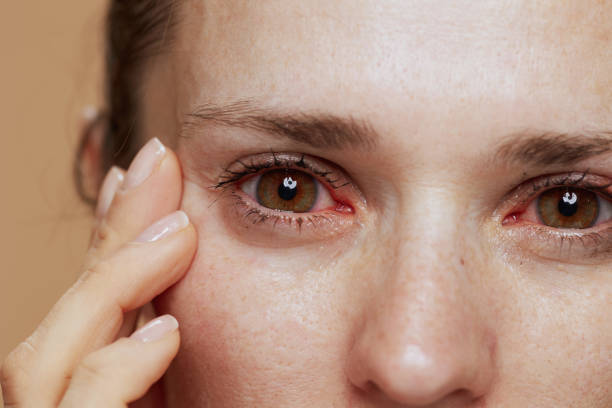
Symptoms Clinical manifestations of dry eye syndrome are very diverse and are largely determined by the severity of the disease. Subjective symptoms of dry eye syndrome include a sensation of a foreign body (sand) in the conjunctival cavity, redness, burning and cutting in the eyes; lacrimation, augmented compassion to light, color blindness, fast fatigue; indistinct vision, pain when filling eye drops.
Symptoms of dry eye syndrome are usually more pronounced in the evening, as well as when in a dry or polluted room, in the cold, wind, or after prolonged or intense visual work.
Objective signs of dry eye syndrome are xerotic changes in the cornea and conjunctiva of varying severity (corneal-conjunctival xerosis). In minor cases of corneal-conjunctival xerosis, compensatory surge in slit manufacture (hyperlacrimia) and an rise in the height of the lower lacrimal meniscus grow.
In moderate xerosis, reflex lacrimation decreases, lacrimal menisci decrease or are completely absent, a feeling of “dryness” in the eyes appears, the swollen conjunctiva creeps onto the free edge of the lower eyelid and shifts along with the stuck eyelid during blinking movements. Severe corneal-conjunctival xerosis is manifested by the following clinical forms: filamentous keratitis , dry keratoconjunctivitis and recurrent corneal erosion, occurring against the background of existing manifestations of eye strain .
In filamentous keratitis, multiple epithelial growths are observed on the cornea, manifestations of moderate corneal syndrome without inflammatory changes in the conjunctiva.
In dry keratoconjunctivitis, pronounced corneal-conjunctival changes of an inflammatory-degenerative nature are observed: subepithelial opacities, dullness and roughness of the cornea, saucer-shaped epithelialized or non-epithelialized depressions on its surface, sluggish hyperemia, edema and forfeiture of gleam of the conjunctiva of the eye, more obvious grip of the look at with the conjunctiva of the eyelids.
With recurrent corneal erosion, superficial microdefects of its epithelium periodically appear, which persist for 3-5 days or more; after their epithelialization, prolonged discomfort is noted.
Particularly severe corneal-conjunctival xerosis usually develops with complete or partial non-closure of the palpebral fissure. Dry eye syndrome against the background of severe vitamin A deficiency manifests itself as squamous metaplasia of the epithelium and keratinization of the conjunctiva.
Dry eye syndrome is often combined with blepharitis . Dry eye syndrome can lead to severe and irreversible xerotic changes and even corneal perforation.
Diagnostics
Diagnostic examination of a patient with dry eye syndrome begins with collecting complaints, assessing the anamnesis and clinical symptoms of the disease, in order to identify pathognomonic and indirect signs of corneal-conjunctival xerosis.
During a physical examination for dry eye syndrome, an external examination is performed, during which the ophthalmologist determines the condition of the skin of the eyelids, the adequacy of their closure, the nature and frequency of blinking movements. During biomicroscopy of the eye , the condition of the tear film, cornea, conjunctiva of the eyeball and eyelids, and the height of the tear meniscus are analyzed.
If dry eye syndrome is suspected, a fluorescein instillation test is performed using a staining solution that allows determining the tear film breakup time and detecting the presence of dry foci – areas of the cornea lacking epithelium. Special tests are used to examine the rate of tear fluid formation – total tear production ( Schirmer test ), the quality and rate of tear film evaporation ( Norn test ). Non-invasive taxation of the precorneal rip picture asset is done consuming tiascopy (examination in polarized light) and measuring the breadth of the wax layer.
A complete ophthalmological examination for dry eye syndrome also includes a laboratory study of osmolarity and crystallography of lacrimal fluid, cytological examination of a smear from the conjunctiva (including impression). In case of systemic or endocrine diseases in the anamnesis of a patient with dry eye syndrome, appropriate immunological and endocrinological studies are carried out.
Treatment of dry eye syndrome
Treatment of dry eye syndrome is aimed at eliminating the etiological factors of xerosis; full moistening of the ocular surface and increasing the stability of the precorneal tear film; stopping pathological changes in the cornea and conjunctiva and preventing complications.
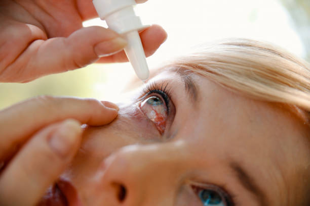
The most widely used method for treating dry eye syndrome is regular instillation of artificial tear preparations (natural tears, gels with carbomer and dexpanthenol), which allow a fairly stable tear film to be restored on the surface of the eyeball. In mild cases of dry eye syndrome, low-viscosity preparations are prescribed, in moderate and severe forms – medium and high viscosity (gels), in especially severe cases of xerosis – low-viscosity preparations without preservatives and also feel like cataracts.
Also, in case of dry eye syndrome, instillations of anti-inflammatory and immunotropic agents are indicated, in the presence of degenerative xerotic changes in the cornea – metabolic drugs. Moreover antihistamines pole cell sheath stabilizers and macrophage lysosomal casing stabilizers are arranged.
Surgery is carried out for dry eye syndrome when steps must be taken to stop the loss and evaporation of tears, boost the supply of tears, eliminate complications (such as dry sores or corneal perforations) and prevent outflow. The closure of lacrimal ducts in the eye may be achieved by using special plugs, conducting plastic surgery at the ducts with conjunctiva or skin or by using diathermocoagulation, laser techniques or surgeries. Obturation of the lacrimal canal with miniature silicone plugs and conjunctival covering of the lacrimal punctum in dry eye syndrome are preferable because they are minimally invasive, more effective and do not cause irreversible changes.
In cases of pronounced corneal xerosis (xerotic ulcer, keratomalacia) and lack of effect from drug therapy and obturation of the lacrimal ducts, keratoplasty is performed in case of dry eye syndrome. Patients with incomplete closure of the eyelids, wide palpebral fissure and rare blinking are indicated for lateral tarsorrhaphy.
Innovative methods of treating dry eye syndrome include transplantation of salivary glands from the oral cavity into the conjunctival cavity, implantation of dacryoreservoirs into the patient’s soft tissues with the introduction of special tubes into the conjunctival cavity.
Prognosis and prevention
Even in mild cases, dry eye syndrome requires comprehensive and adequate treatment to avoid the development of severe diseases of the conjunctiva and cornea with possible loss of vision.
Dry eye syndrome can be prevented by reducing the impact of artificial factors on the eyes, carrying out preventive treatment of internal diseases, including pathology of the visual organs, drinking enough fluids, eating a balanced diet, and blinking more often during visual stress.