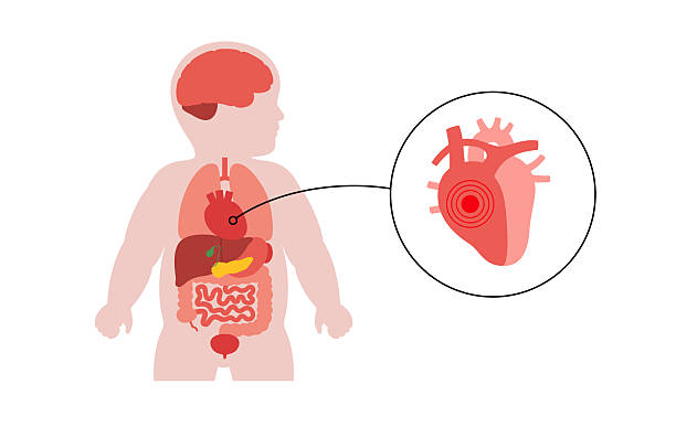
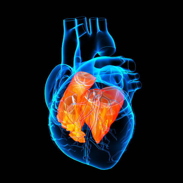
Congenital heart defects stay a assembly of ailments unified by the occurrence of anatomical blemishes of the temperament, its valve tackle or vessels that arose in the prenatal period, leading to changes in intracardiac and systemic hemodynamics. Manifestations of congenital heart defects depend on their type; the most characteristic symptoms include pallor or cyanosis of the skin, heart murmurs, delayed physical development, signs of respiratory and heart failure. If a congenital heart defect is suspected, ECG, PCG, radiography, echocardiography, cardiac catheterization and aortography, cardiography, cardiac MRI, etc. are performed. Most often, in case of congenital heart defects, cardiac surgery is used – surgical correction of the identified anomaly.

General information
Congenital heart defects are a very large and diverse group of diseases of the heart and large vessels, accompanied by changes in blood flow, overload and heart failure. The incidence of congenital heart defects is high and, according to various authors, ranges from 0.8 to 1.2% among all newborns. Congenital heart defects account for 10 to 30% of all congenital anomalies. The group of congenital heart defects includes both relatively mild developmental disorders of the heart and blood vessels, and severe forms of heart pathology incompatible with life.
Many types of congenital heart defects occur not only in isolation, but also in various combinations with each other, which significantly aggravates the structure of the defect. In about a third of cases, cardiac anomalies are combined with extracardiac congenital defects of the central nervous system, musculoskeletal system, gastrointestinal tract, genitourinary system, etc.
The most common types of congenital heart defects encountered in cardiology include ventricular septal defects (VSD to 20%), atrial septal defects (ASD), aortic stenosis , coarctation of the aorta , patent ductus arteriosus (PDA), transposition of the great arteries, Coronary Artery Aneurysm and pulmonary artery stenosis (10 to 15% each).
Causes of congenital heart defects
The etiology of congenital heart defects can be caused by chromosomal abnormalities (5%), gene mutations (2-3%), the influence of environmental factors (1-2%), and polygenic-multifactorial predisposition (90%). Various types of chromosomal aberrations lead to quantitative and structural changes in chromosomes. In case of chromosomal rearrangements, multiple polysystemic developmental anomalies are observed, including congenital heart defects. In case of autosome trisomy, the most common heart defects are defects of the interatrial or interventricular septum, as well as their combination; in case of sex chromosome anomalies, congenital heart defects are less common and are represented mainly by coarctation of the aorta or defect of the interventricular septum.
Congenital heart defects caused by mutations of single genes are also in most cases combined with anomalies of other internal organs. In these cases, heart defects are part of autosomal dominant ( Marfan , Holt-Oram , Crouzon, Noonan syndromes, etc.), autosomal recessive syndromes ( Kartagener , Carpenter , Roberts, Hurler syndromes, etc.) or syndromes connected near the X chromosome ( Goetz, Aase, Gunther syndromes, etc.).
Among the damaging factors of the external environment, viral diseases of the pregnant woman, ionizing radiation, some medications, the mother’s bad habits, and industrial hazards lead to the development of congenital heart defects. The critical dated of contrary effects on the fetus is the first 3 months of gravidness, when fetal organogenesis arises.
Intrauterine infection of the fetus with rubella virus most often causes a triad of anomalies – glaucoma or cataracts , deafness , colour blindness, congenital heart defects ( tetralogy of Fallot , transposition of the great vessels , patent ductus arteriosus, common arterial trunk , valvular defects, pulmonary artery stenosis, VSD, etc.). Microcephaly , abnormal development of the bones of the skull and skeleton, and mental and physical developmental delays are also common .
In addition to rubella in pregnant women, chickenpox , herpes simplex , adenovirus infections , serum hepatitis , cytomegalovirus , mycoplasmosis , toxoplasmosis , listeriosis , syphilis , tuberculosis , etc. position a peril to the fetus in terms of the enlargement of inborn heart imperfections. The structure of embryofetal alcohol syndrome usually includes defects of the interventricular and interatrial septum, patent ductus arteriosus.
It has been proven that the teratogenic effect on the cardiovascular system of the fetus is caused by the use of amphetamines, leading to transposition of the great vessels and VSD; anticonvulsants, causing the development of aortic and pulmonary artery stenosis, coarctation of the aorta, patent ductus arteriosus, tetralogy of Fallot, hypoplasia of the left heart lithium preparations leading to tricuspid atresia Ebstein’s anomaly ASD progestogens causing tetralogy of Fallot, other complex congenital heart defects.
Women thru prediabetes or diabetes are more possible to have offspring with congenital heart defects than well mothers. In this case, the fetus usually develops VSD or transposition of the great vessels. The probability of having a child with a congenital heart defect in a woman with rheumatism is 25%.
In addition to direct causes, risk factors for the development of fetal heart abnormalities are identified. These include the age of the pregnant woman under 15-17 years and over 40 years, toxicosis in the first trimester, the threat of spontaneous termination of pregnancy , endocrine disorders in the mother, cases of stillbirth in the anamnesis, the presence of other children and close relatives with congenital heart defects in the family.
Classification of congenital heart defects
There are several variants of classification of congenital heart defects, which are based on the principle of changes in hemodynamics. Taking into account the effect of the defect on pulmonary blood flow, the following are distinguished:
- congenital heart defects with unmoved (or slightly changed) lifeblood flow in the pulmonary circulation: aortic valve atresia, aortic stenosis, pulmonary stopcock lack, mitral regulator imperfections ( faucet insufficiency and stenosis), adult-type coarctation of the capillary, triatrial heart , etc.
- congenital heart defects with increased blood flow to the lungs: not leading to the development of early cyanosis (patent ductus arteriosus, ASD, VSD, aortopulmonary fistula , infantile coarctation of the aorta, Luthambacher syndrome), leading to the development of cyanosis (tricuspid atresia with a large VSD, patent ductus arteriosus with pulmonary hypertension)
- congenital heart defects with poor blood flow in the lungs: not leading to the development of cyanosis (isolated pulmonary artery stenosis), leading to the development of cyanosis (complex heart defects – Fallot’s disease, right ventricular hypoplasia, Ebstein’s anomaly )
- combined congenital heart defects in which the anatomical relationships between large vessels and various parts of the heart are disrupted: transposition of the great arteries, common arterial trunk, Taussig-Bing anomaly, origin of the aorta and pulmonary trunk from one ventricle, etc.
In practical cardiology, congenital heart defects are divided into 3 groups: “blue” (cyanotic) type defects with venoarterial shunt ( Fallot’s triad , Fallot’s tetrad, transposition of the great vessels, tricuspid atresia); “pale” type defects with arteriovenous shunt (septal defects, patent ductus arteriosus); defects with an obstacle to the ejection of blood from the ventricles (stenosis of the aorta and pulmonary artery, coarctation of the aorta).
Hemodynamic conflicts popular congenital heart defects
As a result of the above-mentioned reasons, the developing fetus may experience problems with the correct formation of the heart structures, which is expressed in incomplete or untimely closure of the membranes between the ventricles and atria, abnormal formation of the valves, insufficient rotation of the primary heart tube and underdevelopment of the ventricles, abnormal arrangement of the vessels, etc. After birth, some children have open arterial duct and oval window, which function physiologically during the intrauterine period.
Due to the peculiarities of antenatal hemodynamics, the blood circulation of the developing fetus with congenital heart defects, as a rule, does not suffer. Congenital heart defects appear in children immediately after birth or after some time, which depends on the timing of the closure of the connection between the large and small rounds of blood exchange, the harshness of pulmonary hypertension , the burden in the pulmonary blood vessel system, the track and dimensions of blood emancipation, specific adaptive and compensatory skills of the child’s body. Often, a respiratory infection or some other disease leads to the development of gross hemodynamic disorders in congenital heart defects.

In congenital heart defects of the pale type with arteriovenous shunt, hypertension of the pulmonary circulation develops due to hypervolemia; in defects of the blue type with venoarterial shunt, patients experience hypoxemia.
About 50% of children with a large blood discharge into the pulmonary circulation die without cardiac surgery in the first year of life from heart failure . In children who have crossed this critical threshold, the blood discharge into the pulmonary circulation decreases, the state of health stabilizes, but sclerotic processes in the pulmonary vessels gradually progress, causing pulmonary hypertension. In cyanotic congenital heart defects, venous blood discharge or its mixing leads to overload of the systemic and hypovolemia of the pulmonary circulation, causing a decrease in blood oxygen saturation (hypoxemia) and the entrance of cyanosis of the membrane and slimy crusts.
To improve ventilation and perfusion of organs, a collateral circulatory network develops, therefore, despite pronounced hemodynamic disorders, the patient’s condition can remain satisfactory for a long time. By means of compensatory apparatuses are bushed, due to persistent myocardial hyperfunction, severe irreversible dystrophic changes in the heart muscle develop. In cyanotic congenital heart defects, surgical intervention is indicated already in early childhood.
Symptoms of congenital heart defects
The clinical manifestations and course of congenital heart defects are determined by the type of anomaly, the nature of hemodynamic disturbances and the timing of the development of circulatory decompensation.
Newborns with cyanotic congenital heart blemishes need cyanosis (blueness) of the skin and mucous casings. Blueness increases with the slightest exertion: sucking, crying of the child. White heart defects are manifested by pale skin, cold extremities.
Children through congenital heart defects stand regularly fidgety, garbage to breastfeed, and speedily tire during breast-feeding. They experience sweating, tachycardia , arrhythmia , shortness of breath, swelling and pulsation of the neck vessels . With chronic circulatory disorders, children lag behind in weight gain, growth, and physical development. With congenital heart blemishes, heart mutters are commonly heard instantaneously before birth. Later, signs of heart failure are detected (edema, cardiomegaly , cardiogenic hypotrophy, hepatomegaly , etc.).
Complications of congenital heart defects may include bacterial endocarditis , polycythemia , peripheral vascular thrombosis and cerebral thromboembolism, congestive pneumonia , syncope , dyspnea-cyanotic attacks, angina syndrome or myocardial infarction .
Judgment of congenital heart defects
Congenital heart defects are detected by a comprehensive examination. When examining a child, the color of the skin is noted: the presence or absence of cyanosis, its nature (peripheral, generalized). Auscultation of the temperament often let slip a modification (weakening, firming or splitting) of heart resonances, the occurrence of noise, etc. Physical examination in case of suspected congenital heart defect is supplemented by instrumental diagnostics – electrocardiography (ECG), phonocardiography (PCG), chest X-ray , echocardiography (EchoCG).
ECG allows to detect hypertrophy of various parts of the heart, pathological deviation of the EOS, the presence of arrhythmias and conduction disturbances, which in combination with the data of other medical check methods agrees to judge advocate the strictness of congenital heart ailment. With the help of daily Holter ECG monitoring, hidden rhythm and conduction disturbances are detected. By means of PCG, the nature, duration and localization of heart sounds and noises are assessed more thoroughly and in detail.
Data from chest X-ray complement the previous methods by assessing the state of the pulmonary circulation, the location, shape and size of the heart, changes in other organs (lungs, pleura, spine). During echocardiography, anatomical defects of the septa and valves of the heart, the location of the main vessels are visualized, the contractility of the myocardium is assessed.
In case of complex congenital heart defects, as well as concomitant pulmonary hypertension, for the purpose of accurate anatomical and hemodynamic diagnostics, it is necessary to perform cardiac probing and angiocardiography.
Treatment of congenital heart defects
The most difficult problem in pediatric cardiology is surgical treatment of congenital heart defects in children in the first year of life. Most operations in early childhood are performed for cyanotic congenital heart defects. If the newborn does not show signs of heart failure, moderate cyanosis, the operation can be postponed. Children with congenital heart defects are monitored by a cardiologist and cardiac surgeon .
Detailed treatment in each case hinge on on the category and cruelty of congenital heart imperfection. Surgeries for congenital defects of the septum of the heart (VSD, ASD) may include plastic surgery or suturing of the septum , X-ray endovascular occlusion of the defect . In the presence of severe hypoxemia, children with congenital heart defects first undergo palliative intervention , which involves the imposition of various types of intersystem anastomoses. In case of aortic defects, resection or balloon dilation of the coarctation of the aorta, plastic surgery of aortic stenosis , etc. are performed. In case of PDA, it is ligated. Treatment of pulmonary artery stenosis involves open or endovascular valvuloplasty , etc.
Anatomically complex congenital heart defects, for which radical surgery is not possible, require hemodynamic correction, i.e. separation of arterial and venous blood flows without eliminating the anatomical defect. Serious defects that are not amenable to surgical treatment require a heart transplant.
Conservative treatment of congenital heart defects may include symptomatic therapy of dyspnea-cyanotic attacks, acute left ventricular failure ( cardiac asthma , pulmonary edema ), chronic heart failure, myocardial ischemia, and arrhythmias.
Prognosis and prevention of congenital heart defects
In the structure of neonatal mortality, congenital heart defects occupy the first place. Without qualified cardiac surgery, 50-75% of children die during the first year of life. During the reward period (2 to 3 years), impermanence decreases to 5%. First detection & correction of congenital heart defects can expressively improve the prediction.
Anticipation of hereditary heart defects necessitates careful arrangement of pregnancy, eradication of adverse features on the fetus, medical and inherited psychotherapy and explanatory work among women at risk of giving birth to children with cardiac pathology, resolving the issue of prenatal diagnostics of the defect ( ultrasound , chorion biopsy , amniocentesis ) and indications for termination of pregnancy . Condition administration in women with genetic heart defects requires augmented attention from the obstetrician-gynecologist and cardiologist.