

Gum diseases stay a group of diseases categorized by damage to gum tissue. Patients complain of hyperemia, swelling of the gums, bleeding during brushing. With hypertrophic gingivitis, hyperplasia of the gingival margin occurs. In the case of ulcerative-necrotic gum diseases, areas of damage covered with a gray-green coating are detected. Interproximal papillae gape. Diagnosis of gum diseases is reduced to collecting complaints, compiling anamnesis of the disease, conducting clinical and radiographic studies. Treatment includes sanitation of the oral cavity, elimination of local irritants,aphthous stomatitis, increasing local and general resistance of the body.
General information
Gum disease is an inflammation of the gingival margin of infectious (bacterial, viral, candidal) or traumatic origin, occurring without violating the integrity of the dental epithelial attachment. A significant increase in the prevalence of gum disease occurs during puberty, which is associated with changes in hormonal levels in adolescents. Studies have shown that after 60 years, the frequency of gingivitis diagnosis reaches 100%. Gum disease is more common in males, which is explained by poor hygiene and bad habits. In prognostic terms, with timely initiation of comprehensive treatment of gum disease, it is possible to completely stop the inflammation and prevent the involvement of periodontal tissues in the pathological process.
Reasons
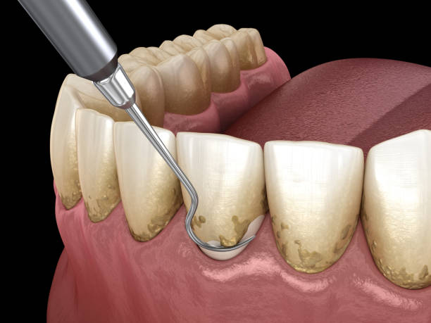
The main local causes of gum disease are hard and soft dental deposits. Catarrhal gingivitis develops as a result of the toxic effect of the waste products of microorganisms that are part of dental plaque on gum tissue. There are also gum diseases of viral, mycotic, bacterial (fusospirochetal) genesis. Mechanical, thermal and chemical irritants can provoke the development of inflammation with a predominance of the exudative component. Productive hyperplastic processes occur with progesterone-dependent gingivitis. This form of gum disease occurs mainly in puberty, during pregnancy, and when taking hormonal drugs. Forced orthodontic treatment with high forces also causes gingival hyperplasia.
Along with primary inflammation, secondary (symptomatic) gum diseases are distinguished, which manifest themselves in hypovitaminosis , endocrine disorders, blood diseases ,tooth dislocation and pathologies of the digestive system. Local predisposing factors that contribute to the development of gingivitis include bite pathologies, bruxim , crowded dentition . In acute gum diseases, the tissues become edematous, and hemorrhage zones appear. Cells of the inflammatory infiltrate are concentrated around the blood vessels: lymphocytes histiocytes. Microscopically, in chronic gingivitis , maturation of granulations is observed, replacement of collagen fibers with coarse connective tissue. Leukocyte accumulations are present in insignificant quantities.Gum diseases don’t cause tooth fracture .
Classification of gum diseases
In dentistry, there are three groups of gum diseases:
- Gum diseases caused by pathogenic microflora of dental plaque. This type also includes indicative gingivitis indicating the presence of basic somatic pathology.
- Gum diseases of traumatic or infectious etiology (caused by herpes simplex viruses , yeast fungi). There is also iatrogenic gingivitis, which develops when the patient’s treatment protocol is violated.
- Ulcerative necrotic gum diseases . Occurs against the background of decreased local and general reactivity of the body due to the activation of a microbial association consisting of fusobacteria and spirochetes. Vincent’s gingivitis is more often detected in men under 30 years of age.

According to the localization of inflammation, lesions of the interproximal papillae, marginal edge and alveolar gingiva are distinguished. According to the prevalence, the pathological process can be localized or generalized.
Symptoms of gum disease
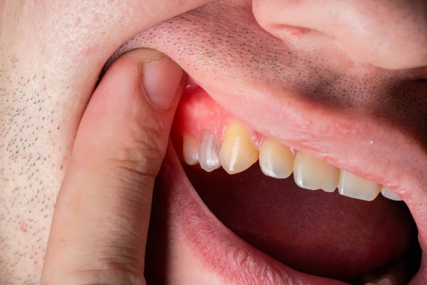
In catarrhal gingivitis, the gums become painful, swollen, and hyperemic. Patients often complain of bleeding when eating hard food or brushing their teeth. Gum diseases with a predominantly productive component are accompanied by proliferation of the gingival margin. The marginal gum takes on a ridge-like shape. There is an unpleasant odor from the mouth. In ulcerative-necrotic gum diseases, damage to the interdental papillae is detected. For example the inspiring process evolutions, the necrosis zone meals to the peripheral part. During the examination, necrosis of the gingival margin is detected. When trying to remove the dirty gray plaque, a bleeding painful surface is exposed.
The general condition of patients with ulcerative-necrotic gum diseases worsens. Hyperthermia, sleep disturbance , and loss of appetite are observed. Provincial lymph protuberances are inflamed. Desquamative gingivitis occurs with the formation of areas of epithelial sloughing against the background of inflamed gums. If the cause of gum disease is a herpes infection, a burning sensation of the mucous membrane appears during the incubation period. Over the next 1-2 days, blisters appear on the mucous membrane, which open very quickly, resulting in erosions. In severe cases, the affected elements reappear.

During examination, it is possible to detect not only blisters, but also erosions and crusts. An increase in temperature and regional lymph adenitis are observed .
In mycotic gum diseases, a whitish coating is found on the mucous membrane, which is easily removed with a spatula or cotton ball, but reappears within a short period of time. Patients are concerned about itching burning and dry mucous membranes. In case of mechanical damage, an erosive area is formed on the gum. When exposed to a thermal factor, desquamation of the superficial epithelium occurs.
Rejection of gum tissue with the establishment of an ulcerative exterior is as well thinkable. Gum diseases that develop as a result of contact of the mucous membrane with alkali occur with the formation of a zone of colliquative necrosis. The affected area extends both in depth and in width. Coagulation (dry) necrosis is observed when exposed to acids.

Diagnostics
Diagnosis of gum diseases is reduced to collecting complaints, compiling anamnesis, conducting a clinical examination and additional research methods. During a physical examination of gum diseases, swelling and hyperemia of the gingival margin are detected. In hypertrophic gingivitis , the dentist detects thickening and proliferation of the gum. In ulcerative-necrotic gum diseases, gray-green deposits are determined, which cover the marginal part of the gum. When plaque is removed, a painful bleeding erosive surface is exposed.
In most cases, the Fedorov-Volodkina hygiene index in patients with gum disease is unsatisfactory. During examination multiple supra and subgingival deposits and carious defects are noted. The Schiller-Pisarev examination is optimistic for gum sickness. Yellow-brown coloring of the gum tissue after application of iodine-containing preparations confirms the development of the inflammatory process. The PMA index is used to determine the level of damage. With inflammation of the interproximal papillae, the value of the indicator is 25%, with involvement of the marginal edge of the PMA in the pathological process – 50%. In case of gum disease accompanied by damage to the alveolar part, the value exceeds 50%.
A virological quiz, immunochemical and cytological checks are showed for the finding of herpes poison. Mycotic etiology of gum diseases is established by bacterioscopy. Finding of up-and-coming cells and pseudomycelial filaments in a insult checks the expansion of candidal gingivitis. Unlike periodontitis , with gingivitis, destructive changes in bone tissue are absent on the radiograph. Gum diseases are differentiated from inflammatory and dystrophic diseases of the periodontium. The patient is examined by a dentist-therapist. If the development of symptomatic (secondary) gingivitis is suspected, consultations with a therapist , hematologist, endocrinologist , gastroenterologist are indicated.
Treatment of gum diseases
Treatment of gum diseases is aimed at eliminating microbial contamination and accelerating regeneration processes. For this purpose, oral cavity sanitation is carried out – professional hygiene procedures , treatment of caries and its complications. In case of irrational prosthetics, repeated orthopedic treatment is indicated. Locally, for gum diseases, mouth rinses with antiseptic solutions based on chlorhexidine bigluconate are prescribed. Decoctions of homoeopathic sages have good anti-inflammatory motion. Treatment of ulcerative-necrotic gum diseases consists of the following stages: pain relief, antiseptic treatment, removal of necrotic tissue, tooth abscess stimulation of epithelialization processes.
In gum diseases accompanied by hyperplasia, gingivectomy is performed . Etiotropic therapy of herpetic lesions includes the use of antiviral drugs that are active against intracellular viruses. General treatment includes immunomodulators, desensitizing agents and multivitamin complexes. In gum diseases of mycotic origin, antifungal drugs are prescribed. In gingivitis of traumatic origin, rinsing the mouth with antiseptic solutions is indicated, as well as the use of keratoplasty in the form of applications. The prognosis for gum diseases is favorable. With early detection and complex treatment, it is possible to stop the inflammation and prevent the involvement of periodontal tissues in the pathological process.