Enamel hypoplasia
Enamel hypoplasia is a dental problem that is accompanied by thinning of the enamel or its complete absence. The uncontrolled complaint can be inborn or developed. Congenital forms are much more common, therefore, children are mostly affected by the disorder. But there are options when teenagers suffer. The disorder leads to a violation of the aesthetics of the dentition, functional problems of various nature. The teeth converted delicate, hurriedly unnatural by caries. Patients require trained behavior under the regulation of a dentist .
Nevertheless you undeniably should not adjournment hypoplasia won’t go away on its own.
Causes of enamel hypoplasia
Enamel hypoplasia is an underdevelopment of the stiff outer sheet of teeth, which is characterized by a stout layer of declared cells. The development is based on various factors of congenital and acquired nature. The pathological condition is most often congenital. It is instigated by the idiosyncrasies of the intrauterine old-fashioned fetal expansion. According to statistics, congenital forms of hypoplasia that affect baby teeth occur in 90% of cases (of the total number of clinical situations). In general, up to 40-50% of the total number of children face the disease. There are fewer teenagers in the incidence statistics, about 10-12% of the general population.
Direct causes of the pathological condition
The formation of pathology most often occurs under the influence of intrauterine factors. Those that affect the child during development.
- severe forms of toxicosis, and the more severe the initial phase of gestation, the higher the likelihood of the child developing dental problems after birth;
- Rhesus conflict, occurs when the fetus has the Rhesus factor and the mother does not have it; such autoimmune conflicts often result in various developmental defects in the child;
- infectious processes suffered during pregnancy, especially dangerous are herpes infections, as well as rubella, measles and some other infections;
- the use of certain medications during pregnancy, hormonal agents in particular are dangerous;
- genetic abnormalities, chromosomal diseases, if the child develops Down syndrome, Marfan syndrome – there is a high probability of parallel development of dental problems of various natures;
- the use of antibiotics, in particular tetracycline, which carries the greatest danger.
Congenital forms of hypoplasia are not the only possible option, however. In about 10% of cases, the Enamel Hypoplasia is acquired.
But it can manifest itself in adolescence. Then the following factors and reasons become provocateurs of pathological processes:
- birth injuries;
- severe diseases of the oral cavity in the period before the change of teeth (for example, advanced caries: the rudiment of a permanent tooth may also be affected);
- use of antibiotics, tetracycline: it affects not only the fetus, but also the person who has already been born;
- Vitamin D deficiency, due to the inability to renew tooth enamel;
- excessive amounts of vitamin D in the body, the defect also appears, but for a different reason: the enamel becomes brittle and quickly wears off, which can also be considered a variant of hypoplasia like bruxism ;
- chemotherapy, chemotherapy drugs suppress regenerative processes, including tooth renewal processes;
- Hormonal imbalance can be both a direct cause and a factor in increasing the risk of disease in a person.
The various associated factors can mostly be corrected.
Pathogenesis of the Enamel Hypoplasia

There are three main mechanisms for the development of the pathological process.
The first is the early death, failure of ameloblast cells. They are responsible for the formation of enamel at the stage of the rudimentary formation. When they die, the tooth is simply not covered with enamel or the process of calcium salt deposition does not end to the extent required to form a strong outer layer of the tooth.
The second mechanism is a deficiency of odontoblast cells. Almost always accompanied by insufficient ameloblast functions. As a result, not only enamel does not form, but also hard tooth structures – dentin, which is even worse.
The third mechanism is a violation of the mineralization process. This is already characteristic of acquired forms of development of the pathological process. There is lacking admission of calcium salts on the now primed base in the form of a protein atmosphere, which is fashioned from unusual cells.
The etiology of enamel hypoplasia is different. But all the described variants are nothing more than theories of the origin of pathology. The exact causes of the pathological process, the lack of a sufficient enamel layer are still unknown. Research continues.
Risk factors for enamel hypoplasia
Risk factors create an increased probability of developing a pathological process in children and adolescents. Among the factors that are found as culprits of a probable congenital anomaly:
- prematurity, in children born before the due date the probability of developing hypoplasia is several times higher;
- severe pregnancy in the mother;
- difficult labor;
- artificial feeding of a child as the basis of nutrition (when the child receives only formula);
- injuries to the maxillofacial region.
If we talk about risk factors for the development of an acquired periodontitis condition in children and adolescents:
- smoking (during adolescence, nicotine affects the rudiments of permanent teeth);
- alcohol consumption
- drug use;
- pronounced hormonal changes, endocrine diseases in the anamnesis;
- poor quality, insufficient nutrition and meager diet;
- history of caries creates risks for permanent teeth.
It is necessary to develop rational preventive measures. Each of the aspects increases the risk by 12-17%.
Classification of enamel hypoplasia
The pathological condition can be divided by which teeth are affected.
Hypoplasia of primary teeth is the most common. It accounts for more than 60% of the total number of clinical cases. It occurs with most of the described changes. In the overwhelming majority of cases, it is due to a severe pregnancy and infectious processes that the expectant mother has suffered.
Hypoplasia of permanent teeth is much less common. It is mainly diagnosed when teeth are replaced by permanent ones. The prerequisites for it are laid in the first 9-12 months of life. It is much less common. Mainly in patients who have suffered severe infectious or metabolic diseases. Less common in patients with endocrine pathology, diseases of the digestive tract. Often, in addition to the described classification method, division by the number of affected teeth is used.
According to our estimates, in 30-40% of cases, patients develop systemic enamel hypoplasia. It is accompanied by damage to all teeth at once. Enamel either develops incompletely or does not develop at all. This type is the most severe. The pathological condition is accompanied by rapid destruction of hard dental tissues with the prospect of their loss. It disturbs together momentary and permanent teeth.
Focal or local enamel hypoplasia is quite rare. In this case, the lesion affects only a few teeth. Usually one or two. Against the background of instrumental damage to the rudiment, carious lesions or other pathological conditions, medical effects. The influence of injuries is also possible. The approach to treating different forms of the Enamel Hypoplasia will be different.
There are other grounds for classifying a violation.
Forms of enamel hypoplasia
Hypoplasia is classified depending on the type of changes, the nature of the underdevelopment of the enamel substance. In this case, the following forms of enamel hypoplasia can be distinguished.
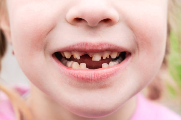
Spotted form – accompanied by the formation of symmetrical spots on the enamel surface. They have a yellowish or white tint. They container remain solitary or multiple, depending on the severity of the condition. Usually do not cause significant discomfort, although they look unaesthetic. The defect is usually superficial, the enamel is partially preserved, as are its functions.
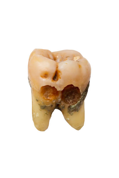
Erosive lesions or cup-shaped hypoplasia are accompanied by the formation of foci of deep enamel hypoplasia. The development of an erosive focus specifies a more forward-thinking form of pathology. Defects are single or multiple.
Grooved hypoplasia is accompanied by the formation of deep lines that run across the tooth, parallel to the cutting edges. Most often, the pathological condition is accompanied by pronounced sensitivity and painful sensations. Enamel hypoplasia progresses quickly.
Mixed forms may occur. In this case, the inferiority of teeth is accompanied by a combination of various forms of enamel structure Enamel Hypoplasia. This is usually the result of complex metabolic disorders. Complex problems with metabolism.
Aplasia stands apart. This is a condition when the tooth germ is formed only partially, incorrectly. There is no enamel at all. In addition to being visually unattractive, the tooth abscess also acquires obvious functional insufficiency. Aplasia is the most severe non-carious lesion of those described.
Types of enamel hypoplasia
Types of hypoplasia are divided depending on the nature of the damage to the teeth.
Hutchinson’s teeth: wounds of the principal points, daggers of the greater &lower jaws each tooth has a conical irregular shape resembling a screwdriver /chisel
- Fournier teeth, in this case the teeth acquire a thickened, barrel-shaped form with a thick neck and a small cutting edge.
A smaller amount common are Pfluger’s teeth: they move the molars. The tooth looks conical. It takes on an irregular shape.
Both type advances for numerous, often not entirely clear reasons. Among other things, different stages of insufficient development of tooth enamel can be distinguished. After birth, teething, the damage to the verbal cavity will only worsen. Until it becomes critical, when it will no longer be possible to save the affected units. The earliest possible start of treatment is indicated.
Symptoms of enamel hypoplasia
Signs of enamel hypoplasia depend on the form and stage of the pathological condition in the patient.
Please note!
The maximum slight are the marked systems of hypoplasia. All the others create a potential threat to the health of temporary and even permanent teeth from the very first moments of development. You cannot delay with the help and consultation of a specialist.
Most often, patients develop the following problems:
- pain: painful sensations are localized in the area of the affected teeth, intensify when chewing, touching &exposed to cold/ hot & sugar;
- increased tooth sensitivity;
- change in the color of the teeth: from white to yellow or yellowish-brown, depending on the situation, which looks unaesthetic;
- changes in the shape and structure of the teeth, they become oval, round or conical, depending on the form of the anomaly;
- the formation of clearly defined visual defects: irregular or regular in shape, often with a clear border (for example, stripes on the front surface of the incisors);
- chewing disorders.
The nature of the symptoms worsens as the pathological process progresses. In case of focal hypoplasia of dental enamel, the clinic affects only the affected units. In case of defects of hard dental tissues, dentin, there is a complete failure of the patient’s dental system. Standard treatment methods are meaningless.
Complications of pathology
Hypoplasia is characterized by a large number of complications. Affected teeth are much less resistant to stress and bacterial factors.
Even with a relatively mild course, patients experience increased and early abrasion of enamel. The pathological condition is accompanied by destruction of the outer layer of the tooth. As a result, the bite is almost always seriously disturbed. Further progression of Enamel Hypoplasia of the dental system is observed.
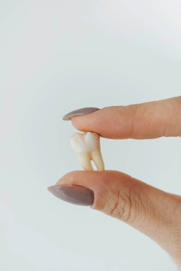
Since the enamel is too weak and fragile, caries develops in the first years of teething. The patient faces pulpitis, tooth fracture and periodontitis, which are dangerous in themselves. Another serious risk is the increased likelihood of damage to the rudiments of permanent teeth. In this case, the problem becomes even more global.
It is almost impossible to avoid the appearance of increased sensitivity of teeth. It becomes difficult to eat. Units affected by the hypoplastic process look unattractive and unaesthetic. And therefore, patients develop psychological problems: isolation, lack of self-confidence.
In some cases, speech disorders develop. Against the background of malocclusion, loss of some teeth. This is not such a rare situation.
Diagnosis of enamel hypoplasia
Hypoplasia of tooth enamel is detected relatively easily. A group of methods is used for diagnostics:
- oral survey to determine complaints and their nature, symptoms of a pathological condition (parents are also often surveyed, since the child cannot accurately formulate complaints);
- collection of anamnesis to assess the probable origin of the pathology;
- visual assessment, examination of the condition of the teeth, as a rule, everything is clear already at this stage;
- X-ray examinations if there are any questions regarding the severity and expression of the pathological process.
The exact list of diagnostic measures is determined by the doctor. It is not difficult to diagnose hypoplasia. However, this is not enough to develop the optimal treatment tactics. It is necessary to understand whether the rudiments of molars have suffered, how much. In what area the foci of hypoplasia have arisen, is this a systemic process or focal. There is a lot of work.
Differential diagnostics of enamel hypoplasia is carried out if there are doubts about the nature of the disorder. The main diseases with which the hypoplastic process is differentiated are fluorosis and caries in the white spot stage. The approaches to therapy will be different. That is why it is important.
Treatment of dental enamel hypoplasia
Treatment depends on a group of factors. The main one is how much the tooth is affected, how deep the enamel defect goes.
When a small number of defects are formed in the form of spots, without symptoms and signs of progression, treatment is usually not required. Dynamic monitoring of the condition is indicated. That is, how the disorder behaves over time.
In relatively mild cases, remineralization is performed, fluoridation is indicated. This is enough to restore, to compensate for the congenital defect. But you need to act quickly, before caries develops.
In case of a deep defect, if a depression appears, thinning of the enamel, but without signs of caries, filling with special materials is carried out. This is important for sealing the tooth. Then – remineralization and fluoridation.
If caries has already developed, it is treated first. But the problem is that the filling in a depleted tooth will not hold. Especially if the delay in enamel development is detected in a milk tooth. Molars are denser. The doctor treats the patient taking into account these restrictions. Then, procedures are also carried out to replenish the enamel deficiency.
In severe forms of damage, when there is no caries, but the enamel layer is too thin and traditional measures are ineffective, treatment of dental enamel hypoplasia is carried out using veneers.
Important!
Veneers are micro-prostheses. Thin plates that allow you to cover the front surface of the teeth from the smile zone. They make the purpose of fake enamel & shielding the teeth. The problem is that the technique is not universal: it allows you to cover only the front teeth. But not the chewing teeth. Crowns are used to correct the condition of the chewing teeth.
When periodontitis foci and gum lesions form, they need to be treated separately. But the situation is still complicated.
Predicting and preventing enamel hypoplasia
The prognosis depends on the type of pathological process, localization and prevalence of the disorder. In most cases, if therapy is started as early as possible, the prognosis is maximally favorable. Moreover, modern dentistry has a large arsenal of correction methods. The worst prognosis is if the teeth are laid down without enamel at all. With aplasia, there is a risk of losing all teeth. In this case, the issue must be resolved using prosthetic methods in two or more stages.
There are no preventive measures developed. But general recommendations can be given. The mother should follow them:
- no smoking;
- do not drink alcohol;
- eat a varied & balanced diet;
- correct hormonal levels;
- avoid infections, and if they develop, treat them immediately;
- avoid stress: preventing emotional and mental overload is very important;
- get enough rest.
Unknown a child matures a problem, you necessity to get a doctor closely. Forward-looking methods of veneer hypoplasia in baby teeth or everlasting teeth lead to adentia. It is possible to restore missing teeth, but this will require much more effort